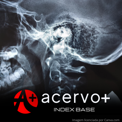
Diferentes métodos de imagem na avaliação da hipertrofia de adenoide
##plugins.themes.bootstrap3.article.main##
Resumo
Objetivo: Analisar sobre o uso da imagem e suas análises qualitativas e quantitativas para diagnóstico da hipertrofia de adenoide. Revisão bibliográfica: A hipertrofia das adenoides causa diversos sintomas, como a respiração oral, roncos e obstrução nasal. As adenoides atingem um pico máximo entre os 3 a 7 anos de idade, a qual depois atrofiam à medida que o paciente envelhece. Atualmente, são diversos os métodos utilizados para a investigação desse quadro, como as imagens bidimensionais estáticas e as tridimensionais dinâmicas. Cada método de diagnóstico apresenta suas vantagens e desvantagens, porém são necessários pois a avaliação clínica é muito subjetiva e inespecífica. Dentre os métodos bidimensionais, tem-se a radiografia de cavum, que fornece informações limitadas de uma região tão complexa, embora muito realizada pelos otorrinolaringologistas. Já os tipos tridimensionais, o mais importante é a videonasolaringoscopia devido à visualização direta de estruturas da nasofaringe. Ainda, a ressonância magnética pode ser utilizada nesses casos devido sua utilidade em diferenciar lesão benigna e maligna. Considerações finais: Devido às características individuais de cada exame e o volume de métodos de análise, é difícil estabelecer qual o melhor exame a ser utilizado, precisando de mais estudos para corroborar ou refutar a literatura existente.
##plugins.themes.bootstrap3.article.details##
Copyright © | Todos os direitos reservados.
A revista detém os direitos autorais exclusivos de publicação deste artigo nos termos da lei 9610/98.
Reprodução parcial
É livre o uso de partes do texto, figuras e questionário do artigo, sendo obrigatória a citação dos autores e revista.
Reprodução total
É expressamente proibida, devendo ser autorizada pela revista.
Referências
2. BALDASSARI CM e CHOI S. Assessing adenoid hypertrophy in children: X-ray or nasal endoscopy? Laryngoscope, 2014; 124(7): 1509-1510.
3. BHATIA KS, et al. Nasopharyngeal mucosa and adenoids: appearance at MR imaging. Radiology. 2012; 263(2): 437-443.
4. CASSANO P, et al. Adenoid tissue rhinopharyngeal obstruction grading based on fiberendoscopic findings: a novel approach to therapeutic management. Int J Pediatr Otorhinolaryngol., 2003; 67(12): 1303-1309.
5. CLEMENS J, et al. Electrocautery versus curette adenoidectomy: comparison of postoperative results. Int J Pediatr Otorhinolaryngol., 1998; 43(2): 115-122.
6. CREPEAU J, et al. Radiographic evaluation of the symptom-producing adenoid. Otolaryngol Head Neck Surg., 1982; 90(5): 548-554.
7. FERES MF, et al. Lateral X-ray view of the skull for the diagnosis of adenoid hypertrophy: a systematic review. Int J Pediatr Otorhinolaryngol. 2011; 75(1): 1-11.
8. FERES MF, et al. Reliability of radiographic parameters in adenoid evaluation. Braz J Otorhinolaryngol., 2012; 78(4): 80-90.
9. FERES MF, et al. Radiographic adenoid evaluation − suggestion of referral parameters. J Pediatr, 2014; 90: 279-85.
10. FUJIOKA M, et al. Radiographic evaluation of adenoidal size in children: adenoidal-nasopharyngeal ratio. AJR Am J Roentgenol., 1979; 133(3): 401-404.
11. KUGELMAN N, et al. Adenoid Obstruction Assessment in Children: Clinical Evaluation Versus Endoscopy and Radiography. Isr Med Assoc J., 2019; 21(6): 376-380.
12. NETO SAA, et al. Avaliação radiográfica da adenóide em crianças: métodos de mensuração e parâmetros da normalidade. Radiol Bras., 2004; 37(6): 445-448.
13. PATINI R, et al. The use of magnetic resonance imaging in the evaluation of upper airway structures in paediatric obstructive sleep apnoea syndrome: a systematic review and meta-analysis. Dentomaxillofac Radiol., 2016; 45(7): 20160136.
14. PARIKH SR, et al. Validation of a new grading system for endoscopic examination of adenoid hypertrophy. Otolaryngol Head Neck Surg., 2006; 135(5): 684-687.
15. PÉREZ-RODRÍGUEZ LM, et al. Airways cephalometric norms from a sample of Caucasian Children. J Clin Exp Dent., 2021; 13(9): e941-e947.
16. SAEDI B, et al. Diagnostic efficacy of different methods in the assessment of adenoid hypertrophy. Am J Otolaryngol., 2011; 32(2): 147-151.
17. SUROV A, et al. MRI of nasopharyngeal adenoid hypertrophy. Neuroradiol J., 2016; 29(5): 408-412.
18. VARGHESE AM, et al. ACE grading-A proposed endoscopic grading system for adenoids and its clinical correlation. Int J Pediatr Otorhinolaryngol., 2016; 83: 155-159.
19. WANG D, et al. Fiberoptic examination of the nasal cavity and nasopharynx in children. Int J Pediatr Otorhinolaryngol., 1992; 24(1): 35-44.
20. WU YP, et al. Hypertrophic adenoids in patients with nasopharyngeal carcinoma: appearance at magnetic resonance imaging before and after treatment. Chin J Cancer, 2015; 34(3): 130-136.

