
Palavras-chave
Periodontia
Tecido Conjuntivo
Implante dentário
Como Citar
Copyright © | Todos os direitos reservados.
A revista detém os direitos autorais exclusivos de publicação deste artigo nos termos da lei 9610/98.
Reprodução parcial
É livre o uso de partes do texto, figuras e questionário do artigo, sendo obrigatória a citação dos autores e revista.
Reprodução total
É expressamente proibida, devendo ser autorizada pela revista.
Resumo
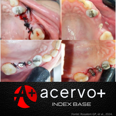
Objetivo: Apresentar um caso clínico de uma reabilitação oral em região de defeito estético com instalação de implantes e uso de enxerto conjuntivo subepitelial para ganho de volume vestibular e tecido queratinizado. Detalhamento de caso: Paciente do gênero feminino, com sessenta anos de idade, se apresentou no dia 11 de outubro de 2019 para fazer a sua primeira consulta clínica no curso de especialização em implantodontia, na Faculdade de Odontologia buscando realizar tratamento reabilitador com implantes. Foi realizada a remoção de tecido gengival livre do túber maxilar, desepitelização e adaptação do enxerto de tecido conjuntivo subepitelial na região vestibular posterior de maxila, juntamente à instalação de dois implantes endósseos. Após o período de osseointegração, provisórios implanto suportados foram confeccionados para adaptação e formação do perfil de emergência peri-implantar. Após a formação da papila interimplantar, as coroas de porcelana foram instaladas. Considerações finais: Considera-se que a técnica combinada de instalação de implantes e inserção de tecido conjuntivo é eficaz para a reabilitação do paciente, nos aspectos funcional e estético.
Referências
BÄUMER D, et al. Socket Shield Technique for immediate implant placement– clinical, radiographic and volumetric data after 5 years. Clinical Oral Implants Research, 2017; 28(11):1450-1458.
BENNANI V, et al. The periodontal restorative interface: esthetic considerations. Periodontology 2000, 2017; 74(1):74-101.
BOYNUEĞRI D, et al. Yeliz A. Significance of keratinized mucosa around dental implants: a prospective comparative study. Clinical oral implants research, 2013; 24(8): 928-933.
CHUNG D, et al. Significance of keratinized mucosa in maintenance of dental implants with different surfaces. Journal of periodontology, 2006;77(8):1410- 1420.
EDEL A. The use of a free connective tissue graft to increase the width of attached gingiva. Oral Surgery, Oral Medicine, Oral Pathology, 1975;39(3):341-346.
FUNATO A, et al. Timing, positioning, and sequential staging in esthetic implant therapy: a four-dimensional perspective. International Journal of Periodontics & Restorative Dentistry, 2007;27(4): 313-323.
GARBER D, et al. Restoration-driven implant placement with restoration-generated site development. Compendium of continuing education in dentistry (Jamesburg, NJ: 1995), 1995;16(8):798-802.
JUNG U, et al. Histologic observation of soft tissue acquired from maxillary tuberosity area for root coverage. Journal of periodontology, 2008; 79(5):934-940.
LANGER B, CALAGNA L. The subepithelial connective tissue graft. The Journal of prosthetic dentistry, 1980;44(4):363-367.
LANGER B, Burton L. Subepithelial connective tissue graft technique for root coverage. Journal of periodontology, 1985;56(12):715-720.
MISCH C. Divisions of available bone in dentistry. Int J Oral Implantol, 1990;7: 9-17.
PAPADOPOULOS I, et al. Transferring the emergence profile from the provisional to the final restoration. Journal of esthetic and restorative dentistry, 2014;26(3): 154-161.
PEREIRA JR, et al. Transferring conditioned partially edentulous ridge form to a master cast. Journal of Prosthodontics, 2016;25(7):595-598.
PUISYS A, LINKEVICIUS T. The influence of mucosal tissue thickening on crestal bone stability around bone‐level implants. A prospective controlled clinical trial. Clinical oral implants research, 2015;26(2):123-129.
REISER G, et al. The subepithelial connective tissue graft palatal donor site: anatomic considerations for surgeons. International Journal of Periodontics & Restorative Dentistry, 1996;16(2):130-137.
SCHUPBACH P, GLAUSER R. The defense architecture of the human periimplant mucosa: a histological study. The Journal of prosthetic dentistry, 2007;97(6): S15-S25.
SON M, JANG H. Gingival recontouring by provisional implant restoration for optimal emergence profile: report of two cases. Journal of periodontal & implant science, 2011;41(6):302-308.
SONICK M. The dependability of connective Tissue Graft-ing for the Resolution of Full-mouth Recession. Compendium, 2011;32(1):48-53.
STEIGMANN M, et al. Emergence profile design based on implant position in the esthetic zone. International Journal of Periodontics & Restorative Dentistry, 2014; 34(4):559-563.
TAN W, et al. A systematic review of post‐extractional alveolar hard and soft tissue dimensional changes in humans. Clinical oral implants research, 2012;23:1- 21.