
Keywords
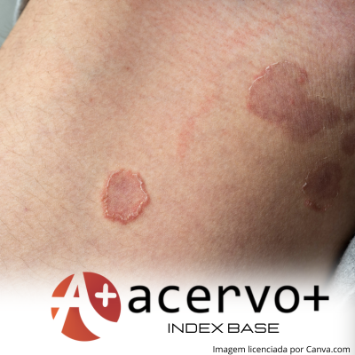
Tineacorporis
Mycology
Dermatology
How to Cite
Copyright © | All rights reserved.
The journal holds the exclusive copyright for the publication of this article under the terms of Brazilian law 9610/98.
Partial reproduction
The use of parts of the texts, figures and questionnaire of the article is free, being mandatory the citation of the authors and journal.
Total reproduction
It is expressly prohibited and must be authorized by the journal.
Abstract
Objective: To analyze the epidemiological andetiological profile of cases of tinea corporis treated at the Specialized Center for Dermatology in a higher education institution in the State of Pará. Methods: This is an observational, cross-sectional study of a descriptive and analytical nature, carried out at the Specialized Center for Dermatology, carried out on patients diagnosed with clinical suspicion of tinea corporis, Who underwent direct mycological examination and culture of the lesions, carried out and analyzed in the dermatology laboratory. Results: 137 individuals with fungal lesions, treated at the afore mentioned location, participated in this study. Of these, 50.4% had tineacorporis. The median age was 55 ± 31 years for tinea corporis and 60.5 ± 23.3 years for the other cases. Considering the general cases of injuries, the majority are female (62%) and with a family income of minimum wage (57.7%). Study limitations: All study parameters could be developed efficiently, with only the culture being performed as the only limiting factor. Conclusion: The study observed a high prevalence of tinea corporis in the city of Belém, Pará, which means that this information and other data collected can be useful in guiding prevention, diagnosis and treatment strategies.
References
AZULAY RD, et al. Dermatologia: Micoses Superficiais. Dermatologia. 6. ed. Rio de janeiro: Guanabara Koogan, 2013; 44: 1188-94.
BALAU ARM. Infeções fúngicas de origem tropical [Dissertação onthe Internet]. [placeunknown]: Instituto Universitário Egas Moniz; 2019; 93.
CANDIDO MB, et al. Experiências vivenciadas em um projeto de pesquisa intitulado “contribuição ao estudo da efetividade e segurança de antifúngicos utilizados para tratamento de micoses superficiais”. Educação, Ciência e Saúde. 2021.
CHANYACHAILERT P, et al. Cutaneous Fungal Infections Caused by Dermatophytes and Non-Dermatophytes: Na Updated Comprehensive Review of Epidemiology, Clinical Presentations, and Diagnostic Testing. J. Fungi, 2023; 9: 669.
COELHO JLG, et al. Dermatófito: resistência a antifúngicos. Brazilian Journal of Development, 2020; 6(10): 74675-74686.
CRUZ CR, et al. Micosis superficiales em la ciudad de Valparaíso, Chile: Período 2007-2009. Rev Chilena Infectol., 2011; 28: 404-9.
DENG R, et al. Dermatophyte infection: from fungal pathogenicity to host immune responses. Front Immunol, 2023; 14: 1-15.
FERRO L de O, et al. Trichophytonrubrum como principal agente etiológico de dermatofitoses em um laboratório de Maceió – AL. Brazilian Journal of health Review, 2020; 3(5): 13198-13207.
GRISOLIA ME. Perfil de sensibilidade aos antifúngicos e de variabilidade genética de espécies de Trichophyton isolados de pacientes com infecção cutânea atendidos em um Serviço Público de Micologia em Manaus/AM [Dissertação onthe Internet]. Manaus: Fundação Oswaldo Cruz Instituto Leônidas e Maria Deane programa de pós-graduação em condições de vida e situações de saúde na Amazônia, 2019; 80.
GUTMANN VLR, et al. Motivos que levam mulheres e homens a buscar as unidades básicas de saúde. J. nurs. Health, 2022; 12(2): e2212220880.
LEUNG AKC, et al. Tinea corporis: an updated review. Drugs in context, 2020; 20: 9.
MELO BLDS, et al. Aspectos epidemiológicos, clínicos e laboratoriais de lesões cutâneas sugestivas de micoses no vale do São Francisco. Brazilian Journal of health Review, 2020; 3(5): 12873-12880.
MEZZARI A, et al. Prevalência de Micoses Superficiais e Cutâneas em Pacientes Atendidos Numa Atividade de Extensão Universitária. Revista Brasileira de Ciências da Saúde, 2018; 21(2): 151-156.
MOSKALUK AE e VANDEWOUDE S. Current Topics in Dermatophyte Classification and Clinical Diagnosis. Pathogens, Fort Collins, 2022; 11.
NEVES IAV. Perfil das dermatofitoses na cidade de Manaus: etiologia e epidemiologia por estudo retrospectivo em 10 anos em um centro de saúde de referência. Fundação Oswaldo Cruz Instituto Leônidas e Maria Deane Programa de pós-graduação em condições de vida e situações de saúde na Amazônia; 2023; 518.
QUATRIN PM, et al. Método convencional para o diagnóstico de micoses: um estudo comparativo. Clinical & Biomedical Research, 2021.
SILVESTRE ECA e QUEIROZ-FERNANDES GM. Fungos dermatófitos e resistência a antifúngicos dermatophytes fungal and antifungal resistance. InterAm J Med Health, 2021; 4: e202101021.
SOARES DM, et al. Tinea capitis: revisão de literatura. Brazilian Journal of Surgery and Clinical Research, 2017; 20(1): 159-163.
TIAGO MRM, et al. Pitiríase versicolor e dermatofitoses diagnosticadas entre os anos de 2006 e 2007 no laboratório de micologia do Instituto Nacional de Pesquisas da Amazônia, Manaus-AM. EsvPuc GO, 2018; 45: 123-129.
YEE G e AL AAM. Tinea Corporis. In: Stat Pearls, 2022.