
Keywords
Maternal-fetal interaction
Placental functions
Placental changes
Fractal method
How to Cite
Copyright © | All rights reserved.
The journal holds the exclusive copyright for the publication of this article under the terms of Brazilian law 9610/98.
Partial reproduction
The use of parts of the texts, figures and questionnaire of the article is free, being mandatory the citation of the authors and journal.
Total reproduction
It is expressly prohibited and must be authorized by the journal.
Abstract
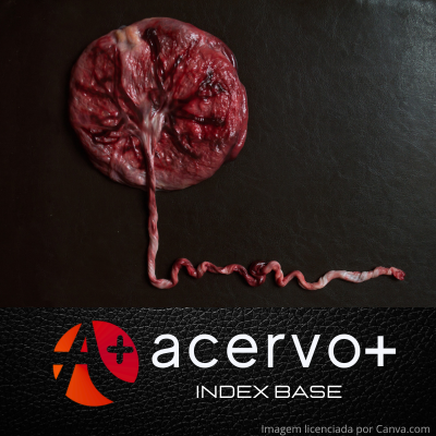
Objective: To analyze the formation, structure and classification for different species and the importance of a well-functioning placenta based on morphological analyses of placental insufficiencies. Bibliographic review: The placenta in mammals is one of the vital structures for maternal-fetal interaction. This structure is responsible for protection against mechanical stress, endocrine regulation during pregnancy, the development of the new individual, as well as facilitating maternal-fetal communication in the exchange of nutritional, gaseous and immunological substances that are fundamental until the birth of the fetus and, if they do not occur as they should, can lead to the death of one or both parties. Therefore, the use of the fractal method to analyze structures helps to detect possible changes. Final considerations: It can be considered that the placenta is very important and has peculiarities for each species in terms of the way it is initially constituted and its internal structures. It was also possible to understand its structure, formation and functions in maintaining maternal and fetal health in humans.
References
BARTHES M, et al. La placenta humana. EMC - Ginecología-Obstetricia, 2025; 61(1): 1-21.
BRÓLIO MP, et al. A barreira placentária e sua função de transferência nutricional. Revista Brasileira de Reprodução Animal, 2010; 34(4): 222-232.
BURTON GJ e JAUNIAUX E. What is the placenta? American Journal of Obstetrics & Gynecology, 2015; 213(4): 1-7.
CARMO LL. Placenta. Kenhub, 2023. Disponível em: https ://www.kenhub.com/pt/library/anatomia/a-placenta. Acesso em: 29 mar. 2025.
CHUCRI TM, et al. Uma revisão da transferência imunológica pela placenta. Revista de Imunologia Reprodutiva, 2010; 87(1-2): 14-20.
CIOBANU AM, et al. Benefits and Risks of IgG Transplacental Transfer. Diagnostics, 2020; 10(8): 583.
COLE LA. Hyperglycosylated hCG, a review. Placenta, 2010; 31(8): 653-664.
Costa DS. Análise fractal e histopatológica de placentas humanas de fetos com restrição de crescimento. Dissertação (Mestrado em Morfotecnologia) – Universidade Federal de Pernambuco, Recife, 2023.
DONNELLY L e CAMPLING G. Functions of the placenta. Anaesthesia & Intensive Care Medicine, 2019; 20(7): 392-396.
FRANDSON RD e WILKE WL, et al. Anatomia e fisiologia dos animais da fazenda. Rio de Janeiro: Guanabara Koogan, 2014; 7: 413.
GRIFFITHS SK e CAMPBELL JP. Placental structure, function and drug transfer. Continuing Education in Anaesthesia Critical Care & Pain, 2015; 15(2): 84-89.
KOTA SK, et al. Endocrinology of parturition. Indian Journal of Endocrinology and Metabolism, 2013; 17(1): 28-35.
MACNAB WR. Functions of the placenta. Anaesthesia and Intensive Care Medicine, 2025; 26(1): 1-5.
MIRON M e MOLDOVANU S, et al. A New Approach in Detectability of Microcalcifications in the Placenta during Pregnancy Using Textural Features and K-Nearest Neighbors Algorithm. Journal of Imaging. 2022; 8(3): 81.
MOORE LK e PERSAUD TVN. Embriologia básica. Rio de Janeiro: Elsevier, 2008; 7: 528.
MOREIRA VR, et al. Aspectos anatômicos da placenta nos animais domésticos. INIC 2024. Universidade Federal do Espírito Santo, 2024.
NOVIS MI, et al. Placental magnetic resonance imaging: normal appearance, anatomical variations, and pathological findings. Radiologia Brasil, 2021.
OLIVEIRA AC. Aspectos da Fisiologia da Placenta nos Animais Domésticos e Retenção de Placenta nos Bovinos. Revisão de Literatura. Monografia (Conclusão do curso de Medicina Veterinária) - Universidade Federal de Campina Grande, Patos, 2015. Disponível em: http ://dspace.sti.ufcg.edu.br:8080/xmlui/bitstream /handle/riufcg /24002/ALINE%20CONCEI%c3%87%c3%83O %20DE%20OLIVEIRA%20%20-TCC%20MED.VETERI N%c3%81RIA%20C STR%202015.pdf?sequence=1&isAllowed =y Acesso: em 28 mar. 2025.
PRADO CVGB, et al. Transporte placentário de gases - Revisão. Revista de Ciência Veterinária e Saúde Pública, 2015; 2(1): 34-44.
RODRÍGUEZ-CORTÉS YM e MENDIETA-ZERÓN H. La placenta como órgano endocrino compartido y su acción en el embarazo normoevolutivo. Revista de Medicina e Investigación, 2014; 2(1): 28-34.
SHAH R e SALAFIA C, et al. Fractal Analysis - Applications and Updates: Placental Growth and Development Analyzed through 2D and 3D Fractals. Intech Open, 2023.
SILVA PC. Impactos do estresse térmico por calor sobre a placenta de cabras: função endócrina e estresse oxidativo. 2022. Tese de Doutorado. (Programa de Pós-Graduação em Zootecnia) - Faculdade de Zootecnia e Engenharia de Alimentos, Universidade de São Paulo, Pirassununga. Disponível em: https ://sucupira-legad o.capes.gov.br/sucu pira/public/consultas/coleta/trabalhoConclusao/viewTrabalhoConclusao.jsf?popup=tru e&id_trabalho=13290164. Ac esso em: 30 abr. 2025.
SKALBA P e DABKOWSKA-HÚC A, et al. The endocrinology of the human placenta. Wiad Lek, 2003; 56(9-10): 475-480.
TURCO MY e MOFFETT A. Development of the human placenta. The Company of Biologists Ltd, 2019.
WOODING FP e BURTON GJ. Comparative placentation: structures, functions and evolution. Berlin: Springer, 2008; 302.
ZAGA-CLAVELLINA V, et al. La prolactina, una hormona que ayuda al feto a protegerse de las infecciones. Con Evidencia, 2025; 6: 4–7.