
Fibroma mole no mamilo: relato de caso
##plugins.themes.bootstrap3.article.sidebar##
##plugins.themes.bootstrap3.article.main##
Resumo
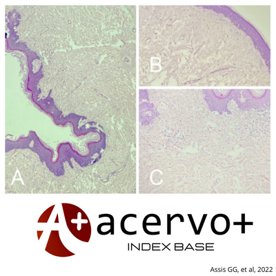
Objetivo: Relatar o caso de uma paciente de 20 anos de idade com apresentação incomum de fibroma mole no mamilo assim como sua excisão cirúrgica com a confirmação do diagnóstico pela histopatologia, revisando também todos os casos já publicados até o momento de fibroma mole no mamilo. Detalhamento do caso: Mulher, 20 anos de idade, com lesão pedunculada, amolecida e indolor no mamilo esquerdo com crescimento insidioso desde seus seis anos de idade. Realizado excisão cirúrgica tangencial e exame histopatológico com achados de acantose irregular, hiperqueratose, papilomatose, derme com tecido conectivo com fibras colágenas frouxas e numerosos capilares com discreto infiltrado mononuclear, evidenciando o diagnóstico de fibroma mole. Considerações finais: O fibroma mole no mamilo é uma lesão benigna rara que geralmente evolui bem após tratamento cirúrgico. Até o momento existem poucas publicações de fibroma mole com esta localização. Mesmo sendo uma neoplasia benigna da pele, o fibroma mole pode acarretar dor local devido a traumas na região, bolhas por atrito local e diminuição da autoestima quando relacionado à questões estéticas e à aceitação pessoal.
##plugins.themes.bootstrap3.article.details##
Copyright © | Todos os direitos reservados.
A revista detém os direitos autorais exclusivos de publicação deste artigo nos termos da lei 9610/98.
Reprodução parcial
É livre o uso de partes do texto, figuras e questionário do artigo, sendo obrigatória a citação dos autores e revista.
Reprodução total
É expressamente proibida, devendo ser autorizada pela revista.
Referências
2. ANSARI MS, et al. Dermoscopy of nipple adenoma. Clin Case Report, 2020; 8:3253–3255.
3. ARORA BK. A clinical case of large fibroepithelial polyp of breast nipple. International Journal of Case Reports and Images, 2019: 10.
4. BANIK R and LUBACH D. Skin tags: localization and frequencies according to sex and age. Dermatologica, 1987; 174: 180-183.
5. BELLI AK, et al. Fibroepithelial Polyp of the Nipple in a Woman. The Breast Journal, 2013; 19: 111-112.
6. BEER T, et al. Tumors of Fibrous Tissue Involving the Skin. In: Elder D, et al. Lever’s Histopathology of the Skin. 11th edition. Philadelphia: Wolters Kluwer; 2015; 2786-2885.
7. COSTA AL, et al. Mammary skin tag in a 2-year-old girl: a long-lasting adnexal polyp of neonatal skin? Pediatric Dermatology; 2009; 26(5): 618-620.
8. DENNIS MA, et al. Incidental Treatment of Nipple Discharge Caused by Benign Intraductal Papilloma Through Diagnostic Mammotome Biopsy. American Journal of Roentgenology. 2000; 174: 1263-1268.
9. ELDER DE, et al. Fibroepithelial polyp. In: Atlas and Synopsis of Lever's Histopathology of the Skin. 2 ed. Philadelphia: Lippincott Williams & Wilkins; 2007: 65–6.
10. ELDER DE, et al. Outline of Cutaneous Pathology. In: Atlas and Synopsis of Lever's Histopathology of the Skin. 2 ed. Philadelphia: Lippincott Williams & Wilkins; 2007: 238-355.
11. GOULD BE, et al. Lack of association between skin tags and colon polyps in a primary care setting. Arch Intern Med. 1988; 148: 1799–800.
12. HIGAKY Y, et al. Blister Formation over a Soft Fibroma of the Nipple. The Journal of Dermatology, 1993; 20: 447-448.
13. HUGHES EJ, et al. Symptomatic fibroepithelial polyp of the nipple in a pediatric patient. Journal of Pediatric Surgery Case Reports, 2022; 76: 102-107.
14. IM B, et al. Imaging Findings of Soft Fibroma of the Nipple: Two Case Reports. J Korean soc Radiol, 2018; 79(3): 123-128.
15. KIM JG, et al. Pyriform-shaped soft fibroma of the nipple in a male patient. The Breast Journal, 2018; 24(5): 827.
16. KIM S and YAUN J. Fibroepithelial polyp of the nipple. Consultant, 2018; 58(1): 46-48.
17. SIM JH, et al. Verrucous epidermal nevus successfully treated with photodynamic therapy. European Journal of Dermatology, 2010; 20: 814-5.
18. MINISTÉRIO DA SAÚDE. Coordenação Nacional de Dermatologia Sanitária. Legislação sobre o Controle de Doenças na Área de Dermatologia Sanitária. Brasília; 1992.
19. NAKAI N, et al. Skin tag of the nipple with blister formation: Two case reports. The Journal of Dermatology, 2013; 40(11): 946-947.
20. NASRI S, et al. Imaging appearance of isolated diffuse neurofibroma of nipple areolar area: a case report. PAMJ, 2021; 39(178).
21. PRIETRO VG and REED RJ. Tumors of Neural Tissue. In: Elder D, et al. Lever’s Histopathology of the Skin. 11th edition. Philadelphia: Wolters Kluwer; 2015. 3188-3189.
22. SEO BF and JUNG S. Fibroma of the Nipple. The Breast Journal, 2012; 18(5): 494-495.
23. SHABAAN AM, et al. An unusual case of a large fibroepithelial stromal polyp presenting as a nipple mass. BMC Research Notes, 2013; 6: 345.
24. SOCIEDADE BRASILEIRA DE DERMATOLOGIA. Perfil nosológico das consultas dermatológicas do Brasil. An Bras Dermatol, 2006; 81: 549-58.
25. VAGHOLKAR K, et al. Pedunculated Fibroma Of The Nipple; Case Report And Review Of The Literature. The Internet Journal of Oncology, 2013; 9: 1-2.
26. VERMA SB. Giant fibroepithelial polyp of the nipple with dermatitis neglecta. J Cutan Aesthet Surg, 2022; 15: 204-206.
27. WALDMAN RA, et al. Skin diseases of the breast and nipple: Benign and malignant tumors. J AM Acad Dermatology, 2019.
28. WALLS SP, et al. Large Fibroepithelial Stromal Polyp of the Breast Nipple. Cureus, 2022; 14(6): e26397.
29. XU X, et al. Diseases Caused by Viruses. In: Elder D, et al. Lever’s Histopathology of the Skin. 11th edition. Philadelphia: Wolters Kluwer; 2015; 1878-1890.
30. YOU HS, et al. A Case of Soft Fibroma of the Nipple with a Cauliflower-Like Appearance. Ann Dermatol, 2015; 27: 102-103.
31. YUCUMÁ D, et al. Soft fibroma of the nipple: Clinical and histopathological characteristics. Rev Senol Patol Mamar, 2020; 34: 60-61.