Tecidos peri-implantares: aspectos histomorfológicos e possíveis apresentações clínicas
##plugins.themes.bootstrap3.article.main##
Resumo
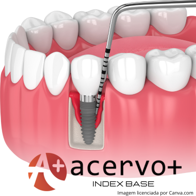
Objetivo: Revisar a temática dos tecidos peri-implantares, seus aspectos histomorfológicos, manifestações clínicas e processos inflamatórios. Revisão bibliográfica: Os tecidos periodontais e peri-implantares apresentam características histomorfológicas semelhantes, embora cada qual apresente particularidades. A saúde peri-implantar caracteriza-se pela presença de tecido conjuntivo coberto por epitélio ceratinizado ou não e sem perda óssea radiograficamente visível. A mucosite peri-implantar é uma condição inflamatória dos tecidos moles ao redor do implante, sem perda óssea além daquela própria à remodelação óssea inicial. A peri-implantite é uma lesão inflamatória da mucosa ao redor de um implante endósseo com perda progressiva do osso peri-implantar de suporte. Considerações finais: Os tecidos periodontais e peri-implantares apresentam características anatômicas e histomorfológicas em comum, embora cada um apresente suas próprias particularidades. Além disso, o conhecimento anatômico e as manifestações clínicas dos processos de saúde e doenças peri-implantares são de extrema importância na prática clínica.
##plugins.themes.bootstrap3.article.details##
Copyright © | Todos os direitos reservados.
A revista detém os direitos autorais exclusivos de publicação deste artigo nos termos da lei 9610/98.
Reprodução parcial
É livre o uso de partes do texto, figuras e questionário do artigo, sendo obrigatória a citação dos autores e revista.
Reprodução total
É expressamente proibida, devendo ser autorizada pela revista.
Referências
2. ALBREKTSSON T e SENNERBY L. State of the art in oral implants. J Clin Periodontol, 1991; 18: 474–481.
3. ARAÚJO M e LUBIANA, NF. Características dos tecidos peri-implantares. Periodontia, 2008; 18: 8-13.
4. ARAUJO MG e LINDHE J. Peri‐implant health. J Clin Periodontol, 2018; 45: 230–236.
5. ARVIDSON K, et al. Histological characteristics of peri-implant mucosa around Branemark and single-crystal sapphire implants. Clin Oral Implants Res, 1996; 7(1): 1-10.
6. BAKAEEN L, et al. The biologic width around titanium implants: histometric analysis of the implantogingival junction around immediately and early loaded implants. Int J Periodontics Restorative Dent, 2009; 29: 297-305.
7. BERGLUNDH T, et al. Morphogenesis of the peri‐implant mucosa: an experimental study in dogs. Clin Oral Implants Res, 2007; 18: 1–8.
8. BERGLUNDH T, et al. Periimplant diseases and conditions: Consensus report of workgroup 4 of the 2017 World Workshop on the Classification of Periodontal and Peri‐Implant Diseases and Conditions. J Clin Periodontol, 2018; 45: 286–291.
9. BERGLUNDH T, et al. Histopathological observations of human periimplantitis lesions. J Clin Periodontol, 2004; 31: 341–347.
10. BERGLUNDH T, et al. The topography of the vasculares systems in the periodontal and periimplant tissues in the dog. J Clin Periodontol, 1994; 21: 189– 193.
11. BERGLUNDH T, et al. Clinical and structural characteristics of periodontal tissues in young and old dogs. J Clin Periodontol, 1991; 18(8): 616-23.
12. BERGLUNDH T e LINDHE J. Dimension of the periimplant mucosa. Biological width revisited. J Clin Periodontol, 1996; 23(10): 971-973.
13. BERGLUNDH T, et al. Morphogenesis of the peri-implant mucosa: an experimental study in dogs. Clin Oral Implant Res, 2007; 18: 1-8.
14. BERGLUNDH T, et al. Protesis tejido-integradas: la oseointegracion en la odontologia clínica. Berlim: Quintesseng Verlags-Gmbh, 1987; 350.
15. CARCUAC O e BERGLUNDH T. Composition of human peri‐implantitis and periodontitis lesions. J Dent Res, 2014; 93: 1083–1088.
16. COCHRAN DL, et al. A prospective multicenter 5‐year radiographic evaluation of crestal bone levels over time in 596 dental implants placed in 192 patients. J Periodontol, 2009; 80: 725–733.
17. DERKS J, et al. Effectiveness of implant therapy analyzed in a Swedish population: early and late implant loss. J Dent Res, 2015; 94: 44‐51.
18. DERKS J, et al. Effectiveness of implant therapy analyzed in a Swedish population: prevalence of peri‐implantitis. J Dent Res, 2016; 95: 43–49.
19. DONLEY TG, et al. Titanium endosseous implant-soft tissue interface: a literature review. J Periodontol, 1991; 153-160.
20. FU JH, et al. Esthetic soft tissue management for theeth and implants. J Evid Based Dent Pract, 2012; 12(3): 129-142.
21. GARCÉS MAS e ESCODA CG. Periimplantitis. Patol Oral Cir Bucal, 2004; 9: 63-74.
22. GARGIULO AW, et al. Dimensions and relations of the dentogingival junction in humans. J Periodontol, 1961; 32(3): 261-267.
23. GUALINI F e BERGLUNDH, T. Immunohistochemical characteristics of inflammatory lesions at implants. J Clin Periodontol, 2003; 30: 14–18.
24. HEITZ-MAYFIELD LJA e SALVI G.E. Peri‐implant mucositis. J Clin Periodontol, 2018; 45(20): 237–245.
25. IVANOVSKI S e LEE R. Comparison of peri-implant and periodontal marginal soft tissues in health and disease. Periodontol, 2018; 76(1): 116-130.
26. JOVANOVIC, SA. Diagnosis and treatment of peri-implant diseases. Curr Opin Periodontol, 1994; 194-204.
27. KAN, JY, et al. Bilaminar subepithelial connective tissue grafts for immediate implant placement and provisionalization in the esthetic zone. J Calif Dent Assoc, 2005; 33(11): 865-871.
28. KLINGE B e MEYLE J. Soft-tissue integration of implants. Consensus report of Working Group 2. Clin Oral Implants Res, 2006; 17: 93–96.
29. LAFAURIE GI, et al. Microbiome and microbial biofilm profiles of peri‐implantitis: a systematic review. J Periodontol, 2017; 88: 1066–1089.
30. LILJENBERG B, et al. Composition of plaque‐associated lesions in the gingiva and the peri‐implant mucosa in partially edentulous subjects. J Clin Periodontol, 1997; 24: 119–123.
31. LINDHE J e BERGLUNDH T. The interface between the mucosa and the implant. Periodontol 2000. 1998; 17: 47–54.
32. LINDHE, J. Clinical Periodontology and Implant Dentistry. 6th ed: Wiley-Blackwell, 2015.
33. LINDHE J e MEYLE J. Peri‐implant diseases: consensus report of the Sixth European Workshop on Periodontology. J Clin Periodontol, 2008; 35: 282–285.
34. MISCH CE. Implante não é dente: comparação com índices periodontais. Prótese sobre Implantes. St Louis: Editora Santos, 2007.
35. MOHENG P e FERIN J. Clinical and biological facts related to oral implant failure: A two-year follow-up study. Implant Dent, 2005; 14: 281-288.
36. MOON IS, et al. The barrier between the keratinized mucosa and the dental implant. An experimental study in the dog. J Clin Periodontol, 1999; 26(10): 658-663.
37. RENVERT S, et al. Peri‐implant health, peri‐implant mucositis, and peri‐implantitis: Case definitions and diagnostic considerations. J Clin Periodontol, 2018; 45(20): 278–285.
38. SCHWARZ F, et al. Peri‐implantitis. J Clin Periodontol, 2018; 45(20): 246–256.
39. SERINO G, et al. Probing at implants with peri‐implantitis and its relation to clinical peri‐implant bone loss. Clin Oral Implants Res, 2013; 24: 91–95.
40. ZITZMANN NU e BERGLUNDH T. Definition and prevalence of peri‐implant diseases. J Clin Periodontol, 2008; 35: 286–291.

